The over 4000 species of decapod shrimps display variations in color that range from bright and colorful to dull and drab, depending on habitat. The function of coloration varies among different groups of animals, including species recognition, mating displays, warning coloration, and camouflage. Visual acuity is notoriously poor in shrimps; the vision of most is best adapted to detection of movement and recognition of general form. As I did research on color in shrimps, it quickly became obvious that its principal function in shrimps is camouflage against visually hunting predators (e.g., fishes and birds), principally disruptive coloration or blending into a background (color mimicry). See Chapter 5 in Bauer (2023) for a review of research on coloration in shrimps.
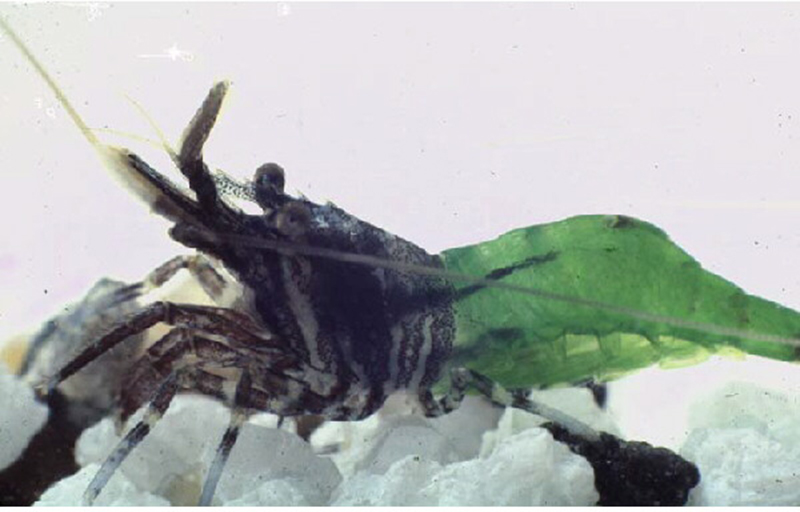
I became interested in studying the biology of shrimps when I first collected the common tidepool shrimp, Heptacarpus sitchensis (formerly H. pictus) at the Bird Rock intertidal area in La Jolla, California. It was hard to believe that the different color forms of this species (photos below), with some individuals mainly a bright "chlorophyll" green, others blotched with the pinks of coralline algae, some with bright white spots similar to bleached shell gravel, could possibly belong to the same species.
|
|
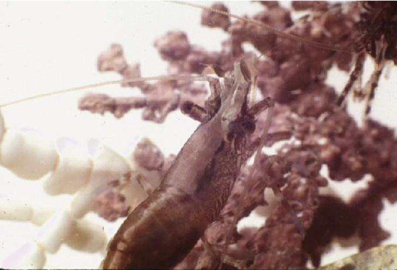
| The "green" morph of Heptacarpus sitchensis, with a banded carapace and a bright green abdomen, similar in color to green algae (Ulva) and the tidepool grass Phyllospadix, both common in the intertidal zone along the west USA coast. | Striped morph with a light pink to reddish middorsal stripe down the length of the body. The stripe is similaar to red corraline algae. |
 |
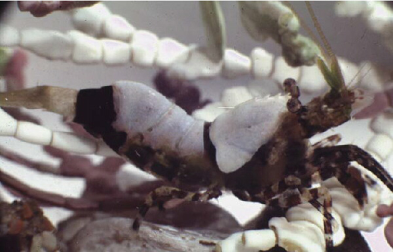 |
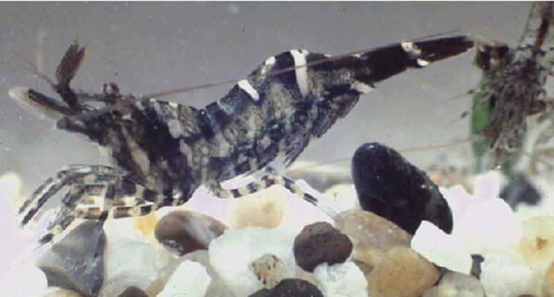
| Banded morph, with dark and white bands and splotches along the body, a good example of disruptive coloration (camouflage) | Color morph of H. taylori, with large splotches of white similar to bleached corraline algae |
The color morphs above are good shrimp examples of disruptive coloration, a form of camouflage in which the bands, stripes and splotches of color distract the eyes of predators from the body profile of the prey, an important visual cue for predators. Disruptive coloration works best against a variable habitat background, such as that of the intertidal zone and shallow sublittoral in which H. sitchensis lives, shown below.
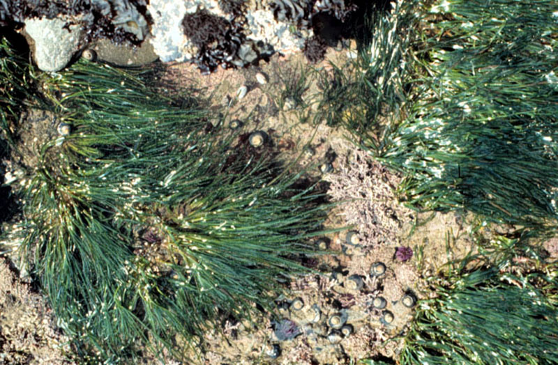 |
| Tidepool of southern California, a color-variable habitat of H. sitchensis with bright green Phyllospadix seagrass, reddish to white (bleached) corraline alage, dark and white gravel, snails etc. |
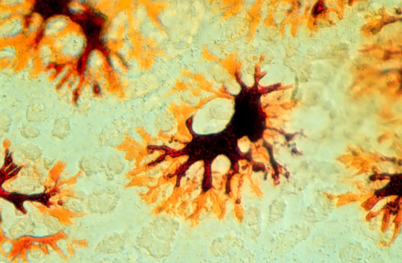
What is the source of color in this and other shrimps? Carotenoid pigments, especially astaxanthin (red) and canthaxanthin (orange, red), are the main source of color in shrimps. When the carotenoids alone produces color, it is red or some shade thereof. When they are linked with proteins (carotenoproteins), colors such as black, brown, violet, blue and green can be produced. When shrimps or other crustaceans with these latter colors are cooked, the carotenoids are unlinked from the denatured proteins, producing the pink to reddish of cooked crustaceans.The bright white "color" is composed of pterin crystals, a noncarotenoid heterocyclic compound, which reflect all wavelengths of visible light (white) to the viewer. The color producing pigments and compounds are either deposited directly into the exoskeleton (cuticle) or contained with color cells (chromatophores) just below the cuticle or deeper within the body. When the cuticle is unpigmented, it is translucent, allowing perception of chromatophores below. Often one or more chromatophores containing the same or different pigments are grouped together into a single structure, termed the "chromatosome." In the older literature, many of the structures termed "chromatophores" are actually chromatosomes (a confusing but important distinction). Chromatophore color is displayed when its pigment is dispersed into the chromatophore's radiating channels; when the pigment is concentrated near the center of the chromatophore, its color is much reduced to the viewer. Below are some examples of the chromatosomes that form color in the caridean Heptacarpus.
 |
 |
 |
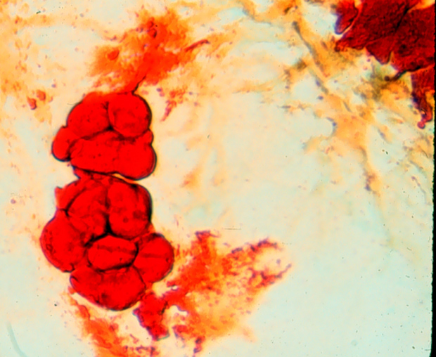
| Red-yellow chromatosome of H. sitchensis, with separate red and yellow chromatophores, their pigments dispersed in cell channels. When in high concentrations, they produce dark bands or splotches. (This and photos to the right were taken with transmitted light through a compound microscope) | Red-yellow chromatophore (higher magnification) , red pigment highly concentrated |
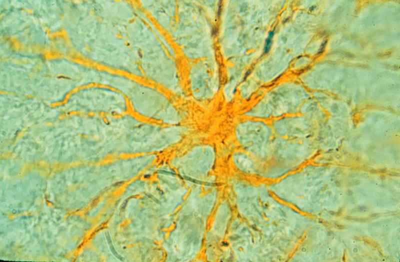
Yellow chromatosome, pigment highly dispersed. When there is a high density of these structures in a body part, the combination of their yellow plus the Tyndall blue of tissues produces the bright green color of the "green morph" when the shrimp is viewed by reflected white light. |
 |
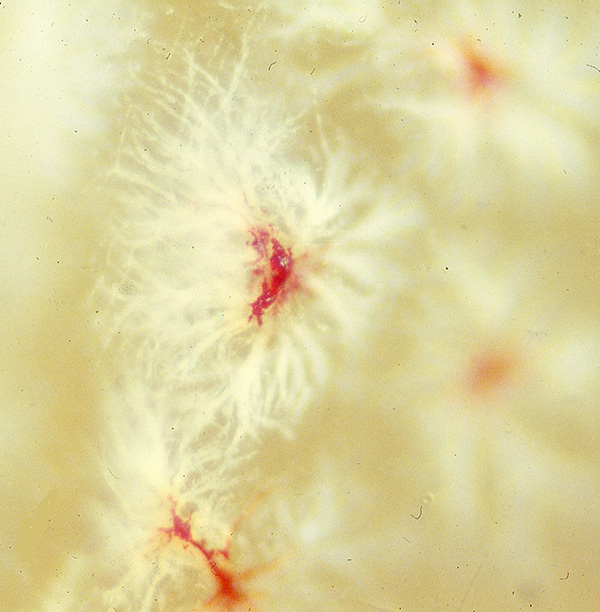 |
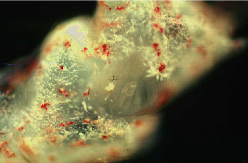
| Red-white chromatosomes in the antennular peduncle, H. sitchensis; in high concentrations, they form a white to pink to reddish color, depending on the dispersion of pigments in the white and red chromatophores that compose the single chromatosomes (viewed with reflected light). | High magnification of a red-white chromatosome, viewed with reflected light. White reflecting substance (probaably crystals of a pterin) are highly dispersed, red pigment is little developed and concentrated in the center of its chromatophore. This type of chromatosome makes up the bright white splotches of color, like that of the H. taylori carapace "saddle" shown above |
Color Change
Many shrimp species are capable of color changes to varying degree. In rapid (physiological) color change, color patterns are displayed with dispersion of pigments within the chromatophores that make up the chromatosomes. When pigments are highly contracted, color fades. "Rapid" color change in shrimps is actually rather slow compared to fast color changes in other aquatic animals such as cephalopods (squids, cuttlefishes, octopi) and some fishes. In cephalopods, the chromatophores are expanded and contracted by muscles fibers under control of the nervous system so that color change is almost instantaneous. In shrimps, dispersion and contraction of pigments are under hormonal control, so that "rapid" color change occurs more slowly, a few minutes up to an hour. In morphological change, which take weeks to occur, the color pattern is changed by the breakdown of chromatosomes and their pigments and replacement with different chromatosomes with different pigments. The stimuli which cause color change is the quality (spectral composition) and intensity of reflected light of the substrate/background color (see Chapter 5 in the Bauer (2023) book on shrimps).
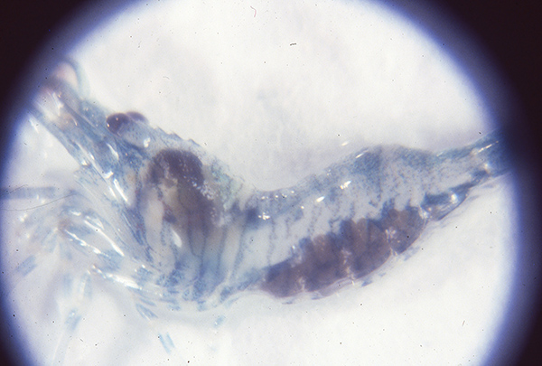
One example of rapid color change is the nocturnal loss of color shown by many shrimp species. I experimented on this phenomenon in the caridean Heptacarpus sitchensis. When laboratory lights were turned off (night), shrimps rapidly loss their bright daytime color, becoming opaque with a bluish tinge (below)
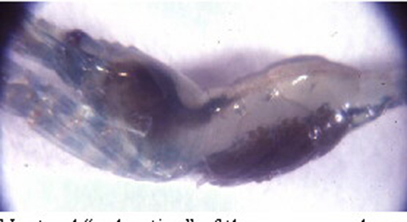 |
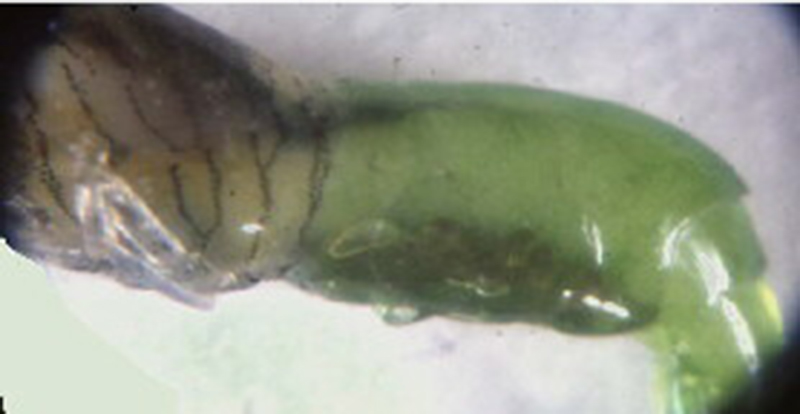 |
| Noctural coloration of the Green color morph of Heptacarpus sitchenis, photo taken in the dark with flash. | Same specimen, 15 minutes after laboratory lights were turned on, green color of abdomen restored |
 |
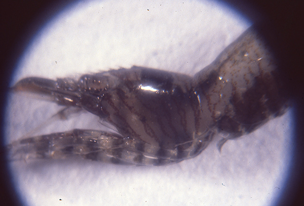 |
| Noctural coloration of the Banded color morph of Heptacarpus sitchenis, photo taken in the dark with flash. | Same specimen, 15 minutes after laboratory lights were turned on, dark color of leg and carapace bands restored, abdomen darker as well. |
One genus of shrimps that are masters of rapid color change in shrimps is Crangon, a benthic shrimp which occurs over sand, gravel and mud substrates. Some species can rapidly adapt their color pattern to match that of the substrate below them, an effective camouflage against visually hunting predators (e.g., fishes, cephalopods, birds).
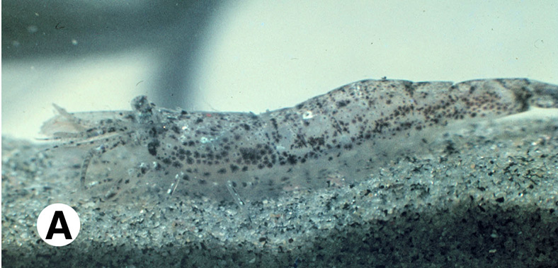 |
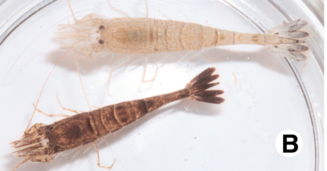 |
| Substrate color matching in a Crangon sp. from Mission Bay (San Diego), California. When viewed from above, the shrimp blends in with its background, an effective camouflage, | Crangon septemspinosa adapted to white (above) and dark (below) backgrounds. The pale specimen above was maintained in a white bucket; the dark-adapted specimen below was transferred to the white bucket just before the photo was taken. |
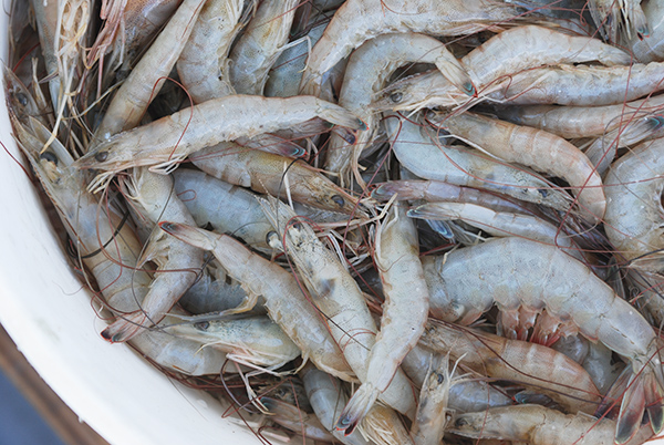
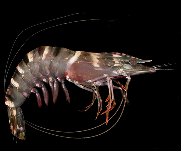
Up to this point, I have been mainly discussing coloration in caridean shrimps, as they (and shallow-water stenopodideans) often display striking color patterns, while benthic penaeoids, which generally occur over drab sand and mud substrates, have correspondingly dull coloration, as does the white shrimp Litopenaeus (Penaeus) setiferus shown below. However, some penaeoids do show stripes and splotches (disruptive coloration; tiger prawn shown below) or bright stripes or "ocelli," that mimic an eye, either startling a potential predator or directing an attack towards its posterior end rather than more essential anterior end.
 |
 |
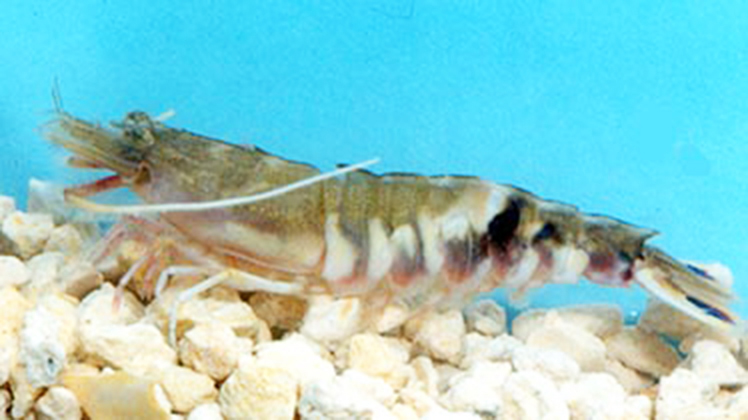 |
| Basket of white shrimp, Litopenaeus setiferus, courtesy of Louisiana Sea Grant | Tiger prawn, Penaeus monodon, courtesy of David Knott, South Carolina Dept. Natural Resources | Sicyonia dorsalis, Gulf of Mexico with white and black bands on the abdomen; some species of Sicyonia have brightly colored "ocelli" (fake eyespots) on the posterior abdomn |
Coloration with Depth
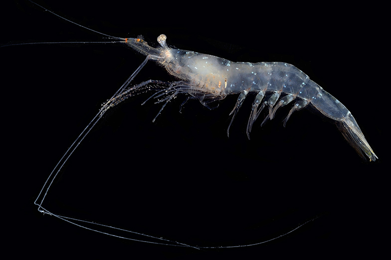
Deep-sea shrimps, both benthic and pelagic, show a reddish coloration with varying degrees of intensity. In pelagic shrimps, coloration ranges from transparent in the upper lighted depths (epipelagic zone, 0-~200 m)), from pale to deeper red in the twilight zone of the mesopelagic zone (~200-1000 m) to deep red at greater depths. These variations with depth are related to camouflage from visually hunting predators such as fishes, birds (epipelagic zone), cephalopods, and marine mammals. The relative glass-like transparency of epipelagic shrimps in the clear lighted waters of the upper open ocean helps hide their bodies from predators. In the dimly lighted waters of the mesopelagic zone, the pale reddish coloration makes them look dark when viewed from above, blending them into the darkness of the waters below. Many of these "half-red" mesopelagic shrimps have evolved light-producing organs (photophores and "Organs of Peseta") which helps hide their "half-red" silhouette when viewed by predators viewing them from below against the light background of the upper lighted waters and sky. Some shrimps can vary their degree of photophore illumination to match ambient lighting as they migrate up to surface waters to feed at night and descend back down as daylight approaches. Please see Chapter 5, Coloration and Camouflage in the Bauer (2023) book on shrimp biology.
 |
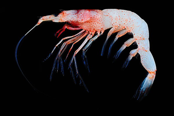 |
| Epipelagic sergestid Sicyonella maldivensis, illustrating transparency of epipelagic shrimps, courtesy of T.-Y. Chan | Challengerosergia talismani, a mesopelagic "half-red" sergestid, courtesy of T.Y. Chan |
 |
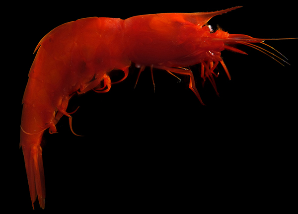 |
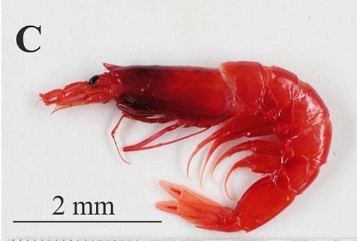
| Robustosergia robusta, a deep mesopelagic sergestid | All-red species Acanthephyra pelagica (Caridea), a bathypelagic aphotic zone |
In the unlighted dark waters of the deep-sea (aphotic) zone, shrimps are invariablyred or some shade thereof. Deep-sea fishes are black in coloration. This led the early deep-sea biologists J. Murray and J. Hjort to term this assemblage of animals as the "red crustacean/prawn, black fish community" in their classic 1912 book "The Depths of the Ocean." Why red shrimps and black fish? In the deep sea, the only source of light which might reveal these animals to predators is bioluminescent light, either that beamed by visually hunting predators themselves or from other organisms. Bioluminescent light is generally in the blue to blue-green wavelengths. We see red objects under white light because the red object reflects red wavelengths to our eyes while absorbing all others. Black objects absorb all wavelengths, white objects reflect all wavelengths. In the deep-sea, red shrimps absorb and do not reflect blue-green light of bioluminescence so that they appear black to a predator and blend in with the black background of deep-sea waters. Fishes do the same using a ground color of black, which is produced by melanin pigments. Shrimps cannot synthesize melanin but they can and do produce the carotenoid astaxanthin, which is red, which in the deep sea produces the same camouflaging effect as does the melanin coloration of deep-sea fishes.
Some deep-sea shrimps, mainly carideans, can emit a luminous cloud of bioluminescent secretion thought to be produced in the heptapancreas, stored in the stomach and emitted through the mouth [Chan, B.K.K., I.-C. Lin, T.-W. Shih and T.-Y. Chan. 2008. Bioluminescent emissions of the deep-water pandalid shrimp, Heterocarpus sibogae De Man, 1917 (Decapoda, Caridea, Pandalidae) under laboratory conditions. Crustaceana 81: 341-350]. Although little studied due the difficulties of collecting and maintaining deep-sea shrimps, it is thought to serve a similar function as the ink cloud emitted by octopi and squids, i.e, to startle or distract an attacking predator. Observations taken via deep-sea submersibles and ROV (Remotely Operated Vehicles) show that the disturbed shrimps swim backwards with tail flips, making their escape behind a bioluminescent screen.
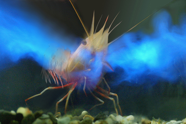 |
| Heterocarpus sibogae, a pandalid deep-sea shrimps emitting a bioluminescent secretion through the mouth (photo courtesy of T.Y. Chan) |
There are many questions to be answered, many opportunities for interesting and important research to be done on these and other aspects of coloration and camouflage in shrimps!
Back to Home Page